| |
Diagnosis of tracheal bronchus in children
Yue-Jie Zheng, Ji-Kui Deng, Dao-Zhen Zhang, Yun-Geng Gen
Shenzhen, China
Author Affiliations: Department of Respiratory Diseases, Shenzhen Children's Hospital, Shenzhen 518026, China (Zheng YJ, Deng JK, Zhang DZ, Gen YG)
Corresponding Author: Yue-Jie Zheng, Shenzhen Children's Hospital, Shenzhen 518026, China (Tel: 86-755-83936199; Fax: 86-755-83936148; Email: yuejiez@sina.com).
Background: Tracheal bronchus is defined as an abnormal bronchus that comes directly off the lateral wall of the trachea above the carina and goes toward the upper-lobe territory. This congenital anomaly may be asymptomatic or present with such symptoms as stridor, cough, wheezing, persistent or recurrent upper-lobe pneumonia, atelectasis or air trapping. This retrospective study was undertaken to summarize the characteristics of tracheal bronchus in children.
Methods: The clinical features of 7 patients with tracheal bronchus were retrospectively analyzed. Of these patients, 2 were initially diagnosed with tracheal bronchus by chest CT and later confirmed bronchoscopically, and 5 by bronchoscopic examination.
Results: Tracheal bronchus was detected in 7 (1.8%) of the 396 patients receiving bronchoscopy. It occurred at the right lateral wall of the trachea in all the 7 patients. Four patients had retained secretions in the tracheal bronchus, which may result in the symptoms of cough, wheezing, and tracheal or bronchial obstruction. Three patients presented with symptoms irrelevant to tracheal bronchus: 1 patient was complicated by laryngomalacia and tetralogy of Fallot, 1 by tracheomalacia, tracheal stenosis and ventricular septal defects, and 1 had mucus plugs in the left lower bronchus.
Conclusion: Tracheal bronchus is a common congenital anomaly in children receiving bronchoscopic examination. More than 50% patients have relevant symptoms. Bronchoscopy is definitely a diagnostic method for tracheal bronchus.
Key words: bronchus; trachea; child; bronchoscopy; abnormality
World J Pediatr 2007;3(4):286-289
Introduction
Tracheal bronchus refers to an abnormal bronchus that comes directly off the lateral wall of the trachea above the carina and goes toward the upper-lobe territory. This uncommon congenital anomaly may be asymptomatic and demonstrated by chest CT or bronchoscopic examination for other respiratory diseases accidentally. It may cause persistent or recurrent upper-lobe pneumonia, atelectasis or air trapping and intubation complications.[1,2] Therefore, understanding tracheal bronchus is of vital importance in clinical management of patients. In this article we summarized the clinical characteristics of 7 children with tracheal bronchus.
Methods
The clinical features of the patients with tracheal bronchus diagnosed bronchoscopically were retro-spectively analyzed. The 7 patients were identified from 396 patients who received rigid or flexible fiberoptic bronchoscopic examination from 2001 to 2006 in Shenzhen Children's Hospital, China. Two patients were initially diagnosed with tracheal bronchus by chest CT, later confirmed bronchoscopically, and 5 patients were diagnosed bronchoscopically. The 7 patients were aged from 4 to 72 months (mean 26 months), and 5 were female and 2 male. All patients presented with symptoms of cough and/or wheezing. Plain chest X-ray showed pneumonia complicated by lower airway obstruction in 2 patients, increased interstitial marking in 4, and right lung hyperinflation in 1. In the 5 patients received chest CT examination, only 2 were diagnosed with tracheal bronchus, 1 with left lung pneumonia complicated by localized hyperinflation, 1 with right bronchus obstruction, and 1 with obstruction at the lower segment of the trachea.
Results
Tracheal bronchus was detected in 7 (1.8%) of the 396 patients receiving bronchoscopy. It occurred at the right side of the trachea in all the 7 patients. Rigid and flexible fiberoptic bronchoscopic image showed anomalous bronchus at the right lateral wall of the trachea above the major carina (Figs. 1, 2). The clinical data of the 7 patients with tracheal bronchus are shown in the Table. Four of the 7 patients (cases 1, 5, 6, 7) had retained secretions in the tracheal bronchus, which was responsible for the symptoms of cough, wheezing, and tracheal or bronchial obstruction. But these symptoms were not attributable to tracheal bronchus in 3 of the 7 patients (cases 2, 3, 4): 1 was complicated by laryngomalacia and tetralogy of Fallot, 1 by tracheomalacia, tracheal stenosis and ventricular septal defects, and 1 had mucus plugs in the left lower bronchus.
Table. Clinical data of the patents with tracheal bronchus
|
Case |
Sex |
Age (mon) |
Compliant |
Chest X-ray |
Chest CT |
Bronchoscopy |
|
1 |
F |
23 |
Cough and wheezing
for 2 days |
Pneumonia, suspected lower airway
obstruction |
Lower airway obstruction, tracheal
bronchus |
Tracheal bronchus accompanied
by retained secretion |
|
2 |
F |
12 |
Cough and fever for
1 week |
Left lung pneumonia accompanied
by localized hyperinflation |
Left lung pneumonia accompanied
by localized hyperinflation |
Mucus plugs of left lower
bronchus, tracheal bronchus |
|
3 |
M |
9 |
Stridor for 8 months,
cough for 3 days |
Increased interstitial marking,
CHD |
Undone
|
Laryngomalacia, tracheal
bronchus |
|
4 |
M |
9 |
Cough and wheezing
for 3 days |
Increased interstitial marking,
CHD |
Undone
|
Tracheomalacia, tracheal
stenosis, tracheal bronchus |
|
5 |
F |
4 |
Cough for 1 week,
wheezing for 1 day |
Increased interstitial marking
|
Lower tracheal stenosis
|
Tracheal bronchus accompanied
by retained secretion |
|
6 |
F |
52 |
Cough and wheezing
for 1 day |
Right lung hyperinflation
|
Right bronchus obstruction |
Tracheal bronchus accompanied
by retained secretion |
|
7 |
F |
72 |
Repeated cough for
2 months |
Increased interstitial marking |
Tracheal bronchus |
Tracheal bronchus accompanied
by retained secretion |
CHD: congenital heart disease; M: male; F: female.
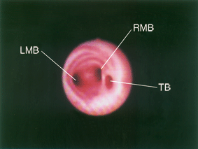
Fig. 1. Bronchoscopy of tracheal bronchus. LMB: left main bronchus; RMB: right main bronchus; TB: tracheal bronchus.
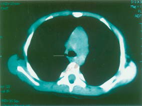
Fig. 2. CT scan of the same patient. CT section demonstrating a right-upper-lobe bronchus (line) arising from the right lateral wall of the trachea above the carina.
Discussion
Tracheal bronchus was first described by Sandifort in 1785 as a right upper bronchus originating from the trachea.[1] The term tracheal bronchus denotes a variety of bronchial anomalies arising in the trachea or main bronchus and directed toward the upper-lobe territory. The reported incidence varies from 1% to 3% in pediatric patients.[1-3] In this study, 1.8% of the patients were detected with the disease by bronchoscopy, suggesting that tracheal bronchus is a common congenital anomalies in children. Tracheal bronchus usually occurs at the right lateral wall of the trachea less than 2 cm above the carina and can supply the entire upper lobe or its apical segment, but it also occurs at the left side of the trachea.[1] There are two types of tracheal bronchus, displaced and supernumerary. If the anatomic upper-lobe bronchus or its single branch is missing, the tracheal bronchus is defined as displaced; displaced type may be the entire upper lobe displaced or its apical segment displaced (high apical lobe). If the right upper-lobe bronchus has a normal trifurcation into apical, posterior, and anterior segmental bronchi, the tracheal bronchus is defined as supernumerary. The supernumerary bronchi may end blindly. In that case, they are also called tracheal diverticula. If they end in aerated or bronchiectatic lung tissue, they are termed apical accessory lungs or tracheal lobes. The displaced type of tracheal bronchus is more frequent than the supernumerary type.[1,4,5] Tracheal bronchus may be complicated by other congenital airway anomalies including laryngomalacia, tracheomalacia, tracheal stenosis, infantile lobar emphysema or associated with congenital heart disease, congenital diaphramatic hernia, Down syndrome and congenital cystic adenomatoid malformation.[6-11] Patients with tracheal bronchus are usually asymptomatic and diagnosed accidentally by bronchoscopy or chest CT for other respiratory diseases. Recent reports including this study indicate that an increasing number of patients with tracheal bronchus present with wheezing, stridor, cough,[12,13] recurrent episodes of infection, hemoptysis, and malignancies.[14-17] In this study 3 patients showed symptoms irrelevant to tracheal bronchus, and the other 4 patients presented with relevant symptoms. In intubated patients, an endotracheal tube was inserted deeply and the tracheal bronchial orifice was obstructed by the tip of the tube, resulting in atelectasis of the involved lobe or segment, post-obstructive pneumonia.[18-22]
Tracheal bronchus may contribute to perioperative persistent hypoxaemia. Thus, tracheal bronchus should be diagnosed in patients with persistent or recurrent upper-lobe pneumonia, atelectasis or air trapping, foreign body aspiration and chronic bronchitis. According to previous reports, most of bronchial branching anomalies are diagnosed by chest CT, especially by techniques of multiplanar reconstruction, three-dimensional reconstruction and three-dimensional virtual bronchoscopy.[1,23-27] In this study, multiplanar reconstruction was not conventionaly done in chest CT, thus only 2 of the 5 patients who had undergone chest CT were diagnosed. This result suggests that the application of CT techniques and understanding of tracheal bronchus are essential to the diagnosis of this disease. With bronchoscopy, the diagnosis could be established by the ectopic bronchus arising from the trachea above the carina. Bronchoscopy is definitely a diagnostic method for tracheal bronchus.
Treatment of tracheal bronchus is based on the severity of symptoms. Most patients with tracheal bronchus can be treated conservatively, but in patients with persistent or recurrent upper-lobe pneumonia, atelectasis or air trapping, surgical excision of the involved segment is necessary.[28]
Funding: None.
Ethical approval: The study was approved by the Ethical Committee of the Shenzhen Children's Hospital, China.
Competing interest: None declared.
Contributors: ZYJ wrote the first draft of this paper. All authors contributed to the intellectual content and approved the final version. ZYJ is the guarantor.
References
1 Berrocal T, Madrid C, Novo S, Gutierrez J, Arjonilla A, Gomez-Leon N. Congenital anomalies of the tracheobronchial tree, lung, and mediastinum: embryology, radiology, and pathology. Radiographics 2003;24:e17.
2 Chau KW, Ng DK, Chong AS, Lau A. Tracheal bronchus. Hong Kong Med J 2003;9:71-72.
3 Aoun NY, Velez E, Kenney LA, Trayner EE. Tracheal bronchus. Respir Care 2004;49:1056-1058.
4 Doolittle AM, Mair EA. Tracheal bronchus: classification, endoscopic analysis, and airway management. Otolaryngol Head Neck Surg 2002;126:240-243.
5 Ghaye B, Szapiro D, Fanchamps JM, Dondelinger RF. Congenital bronchial abnormalities revisited. Radiographics 2001;21:105-119.
6 Keller MS. Congenital lobar emphysema with tracheal bronchus. J Can Assoc Radiol 1983;34:306-307.
7 Elmaci TT, Guler N, Aydogan U, Onursal E. Infantile lobar emphysema and tracheal bronchus in a patient with congenital heart disease. J Pediatr Surg 2001;36:1596-1598.
8 Kairamkonda V, Thorburn K, Sarginson R. Tracheal bronchus associated with VACTERL. Eur J Pediatr 2003;162:165-167.
9 Lee SL, Cheung YF, Leung MP, Ng YK, Tsoi NS. Airway obstruction in children with congenital heart disease: assessment by flexible bronchoscopy. Pediatr Pulmonol 2002;34:304-311.
10 Nose K, Kamata S, Sawai T, Tazuke Y, Usui N, Kawahara H, et al. Airway anomalies in patients with congenital diaphramatic hernia. J Pediatr Surg 2000;35:1562-1565.
11 Bertrand P, Navarro H, Caussade S, Holmgren N, Sanchez I. Airway anomalies in children with Down syndrome: endoscopic findings. Pediatr Pulmonol 2003;36:137-141.
12 McLaughlin FJ, Strieder DJ, Harris GB, Vawter GP, Eraklis AJ. Tracheal bronchus: association with respiratory morbidity in childhood. J Pediatr 1985;106:751-755.
13 Sanchez I, Navarro H, Mendez M, Holmgren N, Caussade S. Clinical characteristics of children with tracheobronchial anomalies. Pediatr Pulmonol 2003;35:288-291.
14 Yildiz H, Ugurel S, Soylu K, Tasar M, Somuncu I. Accessory cardiac bronchus and tracheal bronchus anomalies: CT-bronchoscopy and CT-bronchography findings. Surg Radiol Anat 2006;28:646-649.
15 Kumagae Y, Jinguji M, Tanaka D, Nakajo M. An adult case of bilateral true tracheal bronchi associated with hemoptysis. J Thorac Imaging 2006;21:293-295.
16 Delpizzo KR, Joffe DC, Finkel JC. Tracheal bronchus in 10-month-old patient for thoracoscopic resection of congenital cystic adenomatoid malformation. Paediatr Anaesth 2006;16: 997-998.
17 Vevecka E, De Boeck K, Moerman P, van Raemdonck D, Lerut T. Tracheal bronchus associated with congenital cystic adenomatoid malformation. Pediatr Pulmonol 1995;20: 413-416.
18 O'Sullivan BP, Frassica JJ, Rayder SM. Tracheal bronchus: a cause of prolonged atelectasis in intubated children. Chest 1998;113:537-540.
19 Critchley LA, Ho M, Lee SY. Right upper lobe collapse secondary to an anomalous bronchus after endotracheal intubation for routine surgery. Anaesth Intensive Care 2007; 35:274-277.
20 Wong DT, Kumar A. Case report: endotracheal tube malposition in a patient with a tracheal bronchus. Can J Anaesth 2006;53:810-813.
21 Conacher ID. Implications of a tracheal bronchus for adult anaesthetic practice. Br J Anaesth 2001;86:155-156.
22 Toyoyama H, Minami W, Toyoda Y. Possible right lung isolation by blocking the tracheal bronchus with only a Univent tube for some patients. Anesth Analg 2002;95:492-493.
23 Wong KS, Lien R, Lin TY. Clinical and computed tomographic features of tracheal bronchus in children. J Formos Med Assoc 1999;98:646-648.
24 Shipley RT, McLoud TC, Dedrick CG, Shepard JA. Computed tomography of the tracheal bronchus. J Comput Assist Tomogr 1985;9:53-55.
25 Heyer CM, Kagel T, Lemburg SP, Nicolas V, Rieger CH. Evaluation of tracheobronchial anomalies in children using low-dose multidetector CT: report of a 13-year-old boy with a tracheal bronchus and recurrent pulmonary infections. Pediatr Pulmonol 2004;38:168-173.
26 Newell JD, Thomas HM, Maurer JW. Computed tomographic demonstration of displaced right upper lobe bronchus in an adult woman with congenital heart disease. J Comput Tomogr 1984;8:75-79.
27 Wong KS, Wang CR, Hsieh KH. Demonstration of tracheal bronchus associated with tracheal stenosis using direct coronal computed tomography. Pediatr Pulmonol 1998;25:133-135.
28 Ikeno S, Mitsuhata H, Saito K, Hirabayashi Y, Akazawa S, Kasuda H, et al. Airway management for patients with a tracheal bronchus. Br J Anaesth 1996;76:573-575.
Received February 12, 2007 Accepted after revision June 25, 2007
|
