|
Dermoid cyst of the posterior fossa asso-
ciated with congenital dermal sinus in a child
Chun-Quan Cai, Qing-Jiang Zhang, Xiao-Li Hu, Chun-Xiang Wang
Tianjin, China
Author Affiliations: Departments of Pediatric Neurosurgery (Cai CQ, Zhang QJ), Pathology (Hu XL), and Radiology (Wang CX), Tianjin Children's Hospital, Tianjin 300074, China
Corresponding Author: Chun-Quan Cai, MD, Department of Pediatric Neurosurgery, Tianjin Children's Hospital, Tianjin 300074, China (Tel: +86-22-23519459-86403; Email: tjpns@126.com)
Background: Intracranial dermoid cysts are congenital benign neoplasms. Hydrocephalus and abscess as the principal manifestations of the posterior fossa dermoid cyst are rare. We present a case of obstructive hydrocephalus and abscess induced by an adjacent dermoid cyst with occipital dermal sinus.
Methods: A 2-year-old girl presented with headache and vomiting. Physical examination showed nothing abnormal except for a small subcutaneous nodule above the occipital protuberance with a small skin opening. She had no neurological deficits. Neuroradiological studies including CT and MRI showed a cyst located in the posterior fossa. The cyst in the posterior fossa with occipital dermal sinus was diagnosed. She was treated by radical excision of the occipital cyst through a suboccipital approach, and was followed up.
Results: Histopathologic examination suggested a dermoid cyst with an abscess. Bacterial investigation revealed Staphylococcus epidermidis, and appropriate systemic antibiotic therapy was given. The child recovered and a 2-year follow-up was uneventful.
Conclusions: Posterior fossa dermoid cyst should be considered in all children with occipital skin lesions, especially dermal sinus. CT and MRI scan are helpful in the diagnosis of the lesion. Neurosurgical treatment of the lesion should be planned early to prevent infections such as abscess and meningitis.
Key words: abscess; cranial dermal sinus; dermoid cyst; hydrocephalus; posterior fossa
World J Pediatr 2008;4(1):66-69
Introduction
Intracranial dermoid cyst as congenital benign neoplasm accounts for only 0.1%-0.7% of all intracranial tumors.[1] Clinically, most intracranial dermoid cysts arise in the posterior fossa, a midline position in the vermis or adjacent meninges being favored, and they usually lead to neurological symptoms such as dizziness, headache, and meningitis during childhood. Patients with a posterior fossa dermoid cyst and an associated dermal sinus may develop bacterial meningitis or abscess formation of the dermoid itself.[2] Hydrocephalus and abscess as the principal manifestations of the posterior fossa dermoid cyst are rare. In this report, we present a case of dermoid cyst with contiguous dermal sinus, causing obstructive hydrocephalus and abscess in a girl.
Case report
A 2-year-old girl was admitted to our department, with 20 days history of headache and 4 days of vomiting. The pain was severe at night. She had been born following a normal pregnancy and delivery and with an uneventful period before illness. She denied having any head trauma or neurological deficits.
On admission, physical examination revealed consciousness, a temperature of 37.1ºC, and also a small subcutaneous nodule above the occipital protuberance, which was 4 mm in diameter and had a small skin opening without pus (Fig. 1). The result of routine blood examination was normal. Noncontrast CT of the head showed a well-defined cyst (3 cm ¡Á 3 cm ¡Á 2 cm) located in the midline of the posterior fossa with compression of the adjacent cisterns and the fourth ventricle causing hydrocephalus (Fig. 2). Also, noncontrast MRI of the head showed the similarly well-defined cyst (Figs. 3, 4). Contrast MRI of the head showed the cyst with margin enhancement with compression of the adjacent cisterns and the fourth ventricle causing hydrocephalus (Fig. 5). With the clinical impression of dermoid cyst in the posterior fossa with occipital dermal sinus, suboccipital craniotomy was performed. After opening the dura, a mass containing abscess was noted (Fig. 6). A whitish, midline, encapsulated cystic mass with hair and purulence contents was removed; it was about 3 cm in diameter. The cyst attached to the dura and near the occipital venous sinus bled profusely when it was removed. Histopathologic examination revealed a dermoid cyst (containing lipid and hair) with an abscess of the posterior fossa (Fig. 7). The culture of the discharging material revealed Staphylococcus epidermidis. Followed by systemic antibiotic therapy with ceftriaxone for 2 weeks, the patient's general condition improved rapidly. Post-operative MRI showed the total resection of the lesion with slight edema of the cerebellum and decompression of the adjacent cisterns and the fourth ventricle (Fig. 8). Three weeks after operation, she was discharged without any cerebellar or general neurological symptoms. There is no evidence of recurrence after a follow-up of 2 years, and also no signs of obstructive hydrocephalus at the last visit.
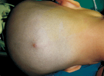 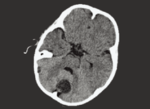
Fig. 1. Occipital small skin opening of the dermal sinus (arrow). Fig. 2. CT showing a well-defined cyst located in the midline of the posterior fossa with compression of the fourth ventricle and dilatation of the lateral ventricle (arrow).
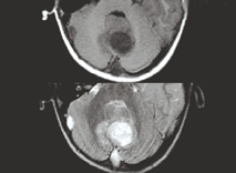 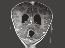
Fig. 3. MRI showing a well-defined cyst located in the midline of the posterior fossa with compression of the adjacent cisterns and the fourth ventricle (arrow). Fig. 4. Contrast MRI showing a well-defined cyst with margin enhancement located in the midline of the posterior fossa and dilatation of the lateral ventricle (arrow).
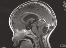 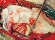
Fig. 5. Contrast MRI showing the margin enhancement of the cyst with compression of the fourth ventricle and dilatation of the lateral ventricle (arrow). Fig. 6. Intra-operation photo showing abscess portion of the mass (arrow).
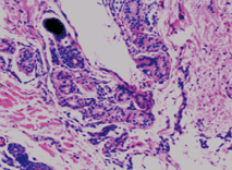 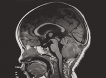
Fig. 7. Histological examination showing dermoid cyst (containing lipid and hair) with purulence contents (HE, original magnification ¡Á 100). Fig. 8. Post-operative MRI showing the total resection of the lesion with slightly edema of the cerebellum and decompression of the adjacent cisterns and the fourth ventricle (arrow).
Discussion
Intracranial dermoid cyst is a congenital benign neoplasm that grows slowly as a result of progressive epithelial desquamation and gland secretion within the cyst. The cyst arises from inclusion of ectodermal elements within the neural tube during its closing between the third and fifth week of embryonic development.[3] It accounts for 0.1%-0.7% of all intracranial tumors,[1] but occurs frequently in the posterior fossa, particularly in the midline position of the vermis or adjacent meninges or in the cavity of the fourth ventricle.[4] Logue and Till[5] reported that dermoid cysts of the posterior fossa tend to lie in the midline of the skull, whether outside or within the dura matter. This tendency to a midline position is probably related to the development of the falx and tentorium, which occurs as an invagination of two folds of the dura, and may draw fragments of the cutaneous ectoderm in with it.[6] Cranial dermal sinuses were first described by Ogle in 1865.[7] Congenital dermal sinuses are congenital dysraphic lesions arising from defective migration of the epithelial ectoderm, postulated to occur through a failure of separation between the cutaneous and neural tube ectoderm. They differ from dermoid and epidermoid tumors in that cranial dermal sinuses have communication with the skin. Although dermoid cyst may rupture and result in aseptic meningitis, cranial dermal sinuses become infected via inoculation of organisms through the sinus tract. This can result in meningitis and abscess formation.[7] Congenital dermal sinuses may occur in the midline anywhere from the nasion to coccyx. The most frequent locations of these sinuses are lumbosacral and occipital regions.[6] Logue and Till[5] classified posterior fossa dermiod cyst into four groups, depending on whether they are extradural or intradural, and on the degree of development of the dermal sinus, whether absent, partial, or complete: (1) an extradural dermoid cyst with a complete dermal sinus, (2) an intradural dermoid cyst without a dermal sinus, (3) an intradural dermoid cyst with an incomplete dermal sinus, and (4) an intradural dermoid cyst with a co12.5n recovery (STIR) images they show low signal intensity. Contrast MRI of the head showed the cyst with margin enhancement, and MRI is of paramount importance to determine the relation of the dermal sinus with venous sinuses. Cerebral angiography usually shows an avascular mass.
Treatment of the posterior fossa dermoid cysts needs microsurgical excision.[9] Total removal of the dermal sinus and the tumor is preferred if permanent cure is ensured. Extreme caution is advised when a sinus is found to penetrate the occipital region. Connection between the dermal sinus, dermoid cyst, and cranial venous confluence is possible, and unanticipated penetration is associated with rapid and fatal exsanguinations. Concerning the infection, we rinsed the bed of the tumor with Amikacin solution to prevent abscess formation post-operation. When hydrocephalus is present, external ventricular drainage promotes more favorable operative conditions and perhaps decreases the likelihood of permanent cerebrospinal fluid diversion.[10] Our patient remained well after surgical excision without previous external ventricular drainage. No recurrence of a posterior fossa dermoid cyst after surgery has been reported in the literature.
In summary, posterior fossa dermoid cyst should be considered in all children with an occipital skin lesion, especially a dermal sinus. Neuroradiological investigations (especially CT and MRI) are helpful in diagnosis. Once diagnosed, microsurgical excision should be performed earlier to prevent the development of severe intracranial complications such as obstructive hydrocephalus, bacterial meningitis and cerebellar abscess.
Funding: No funding.
Ethical approval: Not needed.
Competing interest: None declared.
Contributors: Cai CQ wrote the main body of the article under the supervision of Zhang QJ. Zhang QJ is the guarantor. Hu XL is the helper in the pathological analysis. Wang CX is the helper in the radiological analysis.
References
1 Lunardi P, Missori P, Gagliardi FM, Fortuna A. Dermoid cysts of the posterior cranial fossa in children. Report of nine cases and review of the literature. Surg Neurol 1990;34:39-42.
2 Hsu ST, Lee YY, Chao SC, Hsieh MY, Huang CC. Congenital occipital dermal sinus with intracranial dermoid cyst complicated by recurrent Escherichia coli meningitis. Br J Dermatol 1998;139:922-924.
3 Guidetti B, Gagliardi FM. Epidermoid and dermoid cysts. Clinical evaluation and late surgical results. J Neurosurg 1977;47:12-18.
4 Higashi S, Takinami K, Yamashita J. Occipital dermal sinus associated with dermoid cyst in fourth ventricle. AJNR Am J Neuroradiol 1995;16:945-948.
5 Logue V, Till K. Posterior fossa dermoid cysts with special reference to intracranial infection. J Neurol Neurosurg Psychiatry 1952;15:1-12.
6 Martinez-Lage JF, Ramos J, Puche A, Poza M. Extradural dermoid tumors of the posterior fossa. Arch Dis Child 1997;77:427-430.
7 Aryan HE, Jandial R, Farin A, Chen JC, Granville R, Levy ML. Intradural cranial congenital dermal sinuses: diagnosis and management. Childs Nerv Syst 2006;22:243-247.
8 Akhaddar A, el Hassani MY, Ghadouane M, Hommadi A, Chakir N, Jiddane M, et al. Dermoid cyst of the conus medullaris revealed by chronic urinary retention. Contribution of imaging. J Neuroradiol 1999;26:132-136.
9 Dange N, Bonde V, Goel A, Muzumdar D. Posterior fossa dermoid cyst in a patient with Goldenhar syndrome. Pediatr Neurosurg 2007;43:522-525.
10 Tekkök IH, Baeesa SS, Higgins MJ, Ventureyra EC. Abscedation of posterior fossa dermoid cysts. Childs Nerv Syst 1996;12:318-322.
Received April 26, 2007 Accepted after revision December 4, 2007
|

