|
Diagnosis and management of esophageal
achalasia in children: analysis of 13 cases
Yin Zhang, Chun-Di Xu, Abdehaman Zaouche, Wei Cai
Shanghai, China
Author Affiliations: Department of Pediatrics, Shanghai Ruijin Hospital Affiliated to School of Medicine, Shanghai Jiaotong University, Shanghai 200025, China (Zhang Y, Xu CD); Digestive Functional Exploration Department, Necker Children's Hospital, Paris, France (Zaouche A); Department of Pediatric Surgery, Shanghai Xinhua Hospital Affiliated to School of Medicine, Shanghai Jiaotong University, Shanghai, China (Cai W)
Corresponding Author: Chun-Di Xu, Department of Pediatrics, Shanghai Ruijin Hospital Affiliated to School of Medicine, Shanghai Jiaotong University, Shanghai 200025, China (Tel: +86-21-64370045; Email: chundixu@hotmail.com)
Background: Esophageal achalasia is a rare disease and there have been very few reports about it, especially in children. We reviewed our experience in dealing with esophageal achalasia in 13 children.
Methods: Thirteen children (6 boys and 7 girls), who had been diagnosed with achalasia over a 12-year period between May 1993 and October 2005, were analysed with regard to clinical manifestations, esophageal manometry, endoscopic findings, and treatment. Their age ranged from 3 years to 14 years and 5 months (average 10.3 years) at the time of diagnosis.
Results: In the 13 children, 3 had a family history of esophageal achalasia, 2 of them were sisters. All the 3 children suffered from achalasia/alacrimia/ACTH deficiency. Dysphagia was the most common symptom in the affected children. Vomiting/regurgitation, retrosternal pain, retarded growth, and respiratory symptoms were also observed in some patients. Heller's esophagocardiomyotomy was performed in 9 (69.23%) children, among whom 3 had an antireflux operation at the same time. In the remaining 4 children, 3 received a pneumatic dilatation and 1 received regular administration of nifedipine. Twelve patients were followed up: 8 patients by surgery were cured and have been in perfect condition until now, 3 patients recovered fairly, and 1 patient showed improvement.
Conclusions: Esophageal manometry combined with X-ray examination proves to be an effective diagnostic method for achalasia. It is also effective in evaluating the result of treatment. Heller's esophagocardiomyotomy is a treatment of choice for children with achalasia because of its safety and long-term effective results after surgery.
Key words: dysphagia; esophageal achalasia; esophageal manometry; Heller's esophagocardiomyotomy; pneumatic dilatation
World J Pediatr 2009;5(1):56-59
Introduction
Esophageal achalasia is a rare disease in children and adolescents with unknown cause, and the estimated incidence was 0.11 per 100 000 children.[1] In 1674 Thomas Willis first described this disease as "The mouth of the stomach being always closed, either by a tumor or palsy, nothing could be admitted into the ventricle unless it was violently opened".[2] Regarding the cause of this disease, the most widely accepted theory is a loss of myenteric neurons controlling esophageal smooth muscle.[3] However, "Cruz trypanosomiasis" has been suspected as a possible cause of the disease.[4] Hence it is difficult to prescribe radical treatment for this disease without knowing the definite cause, and the therapeutic effect is difficult to be ascertained.
Reports of this disease are limited because of its extraordinary low incidence in children especially in China where documented cases are rare. We analyzed some documented cases of this disease, and hope the results of this analysis can serve as a useful reference in the diagnosis of esophageal achalasia in our practice.
Methods
Between May 1993 and October 2005, 13 children with achalasia were diagnosed in Shanghai Ruijin Hospital and Necker Children's Hospital of Paris, including 6 boys and 7 girls with an average age of 10.3 years (range: 3-14.42 years). Among them, two were diagnosed at <5 years old, three between 5 and 10 years old, and eight >10 years. The duration of the disease before the diagnosis varied from 3 to 108 months with an average of 31 months.
The diagnosis of achalasia was based on clinical symptoms of one or more of the following presentations: dysphagia, vomiting/regurgitation of food, retrosternal pain, poor growth and respiratory symptoms. An esophageal manometry was done in some patients to confirm the diagnosis. Gastroscopy, pH measurement and barium study were also done in the majority of the patients.
Achalasia was principally diagnosed by clinical symptoms (especially dysphagia), barium meal examination and esophageal manometry.
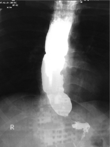
A "bird beak" was usually seen at the junction of the esophagus and stomach with dilation of the esophageal body and absence of esophageal peristalsis. Esophageal manometry was used to measure the pressure of the lower esophageal sphincter (LES) and the superior esophageal sphincter (SES). Deglutitory relaxation was absent or incomplete. The amplitude of peristaltic contraction of the esophageal body was also visualized. The criteria of esophageal manometry for diagnosing achalasia included LES pressure >normal (22¡À10 mmHg), partial or complete lack of relaxation (relaxation rate <90%), abnormal esophageal peristalsis and/or an inversion of the gastroesophageal gradient. Barium swallow showed a "bird beak" at the level of gastroesophageal junction in an achalasia patient (Fig. 1).
The majority of the patients were treated surgically and by pneumatic dilation. In a long-term follow-up, we evaluated the results of treatment by four grades: perfect (no symptoms and check-up studies after treatment showed normal results), good (few mild symptoms and examinations showed some abnormal results), improved (symptoms and results of examinations were better than those found before) and unchanged.
Results
Demographics
In the 13 patients, 6 boys and 7 girls, there was no significant difference in mean age between boys and girls (P=0.168). The average age was 10.3 years (range: 3-14.42 years) at the time of diagnosis. Three patients had a family history of esophageal achalasia and achalasia/alacrimia/ACTH deficiency (AAA) syndrome, 2 of whom were siblings with dysautonomia and the other patient had a sister with AAA syndrome.
Clinical symptoms
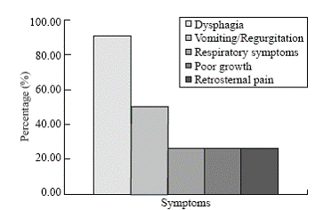
Dysphagia was seen in 10 (92.31%) of the patients, vomiting/regurgitation of food in 7 (53.85%), poor growth in 3 (23.08%), respiratory symptoms in 3 (23.08%) and retrosternal pain in 3 (23.08%) (Fig. 2).
Diagnosis
Esophageal manometry was performed in 11 patients. Incomplete relaxation of the LES and abnormal esophageal peristalsis were seen in all the patients. Ten of the 11 patients showed an increased pressure of the LES (mean: 53.98 mmHg) and an inversion of the gastroesophageal gradient. Abnormal motility of the SES was also seen in 3 patients, showing an incomplete relaxation and increased pressure of the SES (Table).
Barium meal examination in all patients showed diagnostic signs of achalasia: an esophageal dilation and a "bird beak" at the cardia. Esophago-gastroduodendoscopy performed in 8 patients showed dilated esophagus in 3 patients and esophagitis in 1 patient. 24-hour pH measurement in 4 patients showed normal results.
Treatment and follow-up
Most patients were treated surgically to reduce the pressure of the LES. Nine patients received standardized Heller's esophagomyotomy, and their esophageal hiatus was fully dissected for subsequent myotomy.[5,6] Among them, 3 patients also received NISSEN antireflux operation, and only one patient had to undergo Heller's esophagomyotomy for a second time because of failure of the first operation. For the remaining 4 patients, 2 underwent pneumatic dilation twice. One of the two siblings remained clinically stable after the medical treatment with nifedipine, and the other was lost to follow-up after endoscopic dilatation.
The mean follow-up time was 15.2 months (range: 2 months to 4 years). The outcome was "perfect" in 8 patients (66.67%) without any symptoms, including 6 patients by surgery and 2 patients by pneumatic dilatation. The other 3 patients by surgery who had not received antireflux operation were evaluated as "good", one of whom had residual achalasia (dysphagia) and 2 developed gastroesophageal reflux disease. The only patient who had not received surgical treatment showed improvement of symptoms after medication.
 
Fig. 1. Barium swallow of an 8-year-old girl with achalasia demonstra-ting distal esophageal dilatation and narrowing at the lower esophageal sphincter ('bird beak' deformity). Fig. 2. Major symptoms of esophageal achalasia.
Table. Results of esophageal manometry in 11 patients
|
Variables |
No. of patients (%) |
|
Lower esophageal sphincter |
|
Increased pressure |
10 (90.91), mean: 53.98 mmHg |
|
Incomplete relaxation |
11 (100.00) |
|
Abnormal esophageal peristalsis |
11 (100.00) |
|
Inversion of gastroesophageal gradient |
10 (90.91) |
|
Superior esophageal sphincter |
|
|
Increased pressure |
2 (18.18) |
|
Incomplete relaxation |
1 (9.09) |
|
Normal |
8 (72.73) |
Discussion
The major manifestation of esophageal achalasia is the disorder of esophageal motility. The cause of this disease is unknown. The most commonly accepted cause is a pathological change in the nervous system, not the muscle. Anatomical study discovered that loss or absence of nerve cells in the nervous system of esophageal muscle is the major pathological change of this disease. This disease can be seen in any age or race without a clear hereditary trend.[7] It rarely occurs in infants, only 6% of the reported cases in childhood are found in infancy at the time of diagnosis.[8] None of the infants was found in the present series, and the youngest patient was 3 years old. Different studies showed different gender distribution.[9-11] Some reported there is no difference in gender, which is true in the present study. Dysphagia is the most significant symptom in this disease, and symptoms could be triggered or aggravated by swallowing. In this study 3 patients had typical AAA symptoms, i.e., achalasia, alacrimia and ACTH deficiency. They were girls from two unrelated families, 2 of whom were sisters. The sisters had similar symptoms and treatment results. Though the role of heredity was not determined, patients in this study did indicate some trends in heredity.[12,13] Further study in more cases is necessary in order to elucidate this role.
There are several methods for a definite diagnosis of esophageal achalasia. Barium meal examination of the esophagus is the oldest and commonly used method which is very effective in early determination of the disease. Gastroendoscopy is necessary for patients with dysphagia and retrosternal pain to rule out other esophageal pathology, especially esophagitis or tumor of the cardia because clinical symptoms, X-ray findings and manometric test results are very similar to those of achalasia. At present, esophageal manometry has been widely used in the diagnosis of esophageal achalasia because of its simple manipulation and high accuracy.[14] The pressure curve is very accurate and sensitive in detecting basic pressure of different parts of the upper digestive tract and their changes during swallowing. The pressure could be a good indicator for the severity of the disease, for selection of proper treatment and for evaluation of treatment results. Besides 2 cases in our series, the other 11 patients were diagnosed by pressure tests. After treatment, all patients showed normal results of their tests.
In the treatment of this disease, reduction of the pressure of the LES is the primary objective. As a non-surgical treatment, pneumatic dilatation is a traditional and effective treatment.[15] Since the 1970s, it has been used to treat adult patients and children in some hospitals.[16] Compared with esophagomyotomy, this treatment is effective, but costs less and is easily manipulated. It can be used as a treatment of choice for achalasia in children in China. But this treatment has some shortcomings like a relatively short effective period and re-treatment. Two of 3 patients in our study who had received this treatment had to be re-treated. Heller's esophagocardiomyotomy invented in 1913[17] was popularized by Zaaijer around 1923.[18] The treatment has a high degree of safety, effective results, and a long effective period.[19,20] It has been a standard surgical treatment for achalasia in North America and Europe in the resent years. But this treatment may result in some postoperative sequelae such as esophageal reflux. In order to minimize this undesirable effect, this operation is usually performed in combination with antireflux operation and it has become a routine procedure in some countries.
Funding: None.
Ethical approval: Not needed.
Competing interest: None declared.
Contributors: Zhang Y wrote the main body of the artical under the supervison of Xu CD. Zaouche Z and Cai W provided advice on medical aspects. Xu CD is the guarantor.
References
1 Mayberry JF, Mayell MJ. Epidemiological study of achalasia in children. Gut 1988;29:90-93.
2 Willis T. The companation of madication and surgery of esophageal achalasia. In: Willis T, eds. Achalasia. London: Hgas-comitis, 1674: 18-19.
3 Goldblum JR, Rice TW, Richter JE. Histopathologic features in esophagomyotomy specimens from patients with achalasia. Gastroenterology 1996;111:648-654.
4 Kahrilas PJ. Esophageal motility disorders: current concepts of pathogenesis and treatment. Can J Gastroenterol 2000;14:221-231.
5 Smith CD, Stival A, Howell DL, Swafford V. Endoscopic therapy for achalasia before Heller myotomy results in worse outcomes than heller myotomy alone. Ann Surg 2006;243:579-584.
6 Hunter JG, Trus TL, Branum GD, Waring JP. Laparoscopic Heller myotomy and fundoplication for achalasia. Ann Surg 1997;225:655-664.
7 Asch MJ, Liebman W, Lachman RS. Esophageal achalasia: diagnosis and cardiomyotomy in a newborn infant. J Pediatr Surg 1974;9:911-915.
8 Myers NA, Jolley SG, Taylor R. Achalasia of the cardia in children: a worldwide survey. J Pediatr Surg 1994;29:1375-1379.
9 Jara FM, Toledo-Pereyra LH, Lewis JW, Magilligan DJ Jr. Long-term results of esophagomyotomy for achalasia of esophagus. Arch Surg 1979;114:935-936.
10 Okike N, Payne WS, Neufeld DM, Bernatz PE, Pairolero PC, Sanderson DR. Esophagomyotomy versus forceful dilation for achalasia of the esophagus: results in 899 patients. Ann Thorac Surg 1979;28:119-125.
11 Hussain SZ, Thomas R, Tolia V. A review of achalasia in 33 children. Dig Dis Sci 2002;47:2538-2543.
12 Mukhopadhya A, Danda S, Huebner A, Chacko A. Mutations of the AAAS gene in an Indian family with Allgrove's syndrome. World J Gastroenterol 2006;12:4764-4766.
13 Sellami D, Bouacida W, Frikha F. Allgrove syndrome. Report on a family. J Fr Ophtalmol 2006;29:4184-4221.
14 Ragunath K, Williams JG. A review of oesophageal manometry testing in a district general hospital. Postgraol Med J 2002;78:34-36.
15 Mehta R, John A, Sadasivan S, Mustafa CP, Nandkumar R, Raj VV, et al. Factors determining successful outcome following pneumatic balloon dilation in achalasia cardia. Indian J Gastroenterol 2005;24:243-245.
16 Boyle JT, Cohen S, Watkins JB. Successful treatment of achalasia in childhood by pneumatic dilation. J Pediatr 1981;99:35-40.
17 Heller E. Extramukose cardioplastik beim chronischen cardiospasmus mit dilation des esophagus. Mitt Grenzgeb Med Chir 1914;27:141.
18 Zaaijer J. Cardiospasm in the aged. Ann Surg 1923;77:615-617.
19 Ortiz A, de Haro LF, Parrilla P, Lage A, Perez D, Munitiz V, et al. Very long-term objective evaluation of heller myotomy plus posterior partial fundoplication in patients with achalasia of the cardia. Ann Surg 2008;247:258-264.
20 Bonavina L. Minimally invasive surgery for esophageal achalasia. World J Gastroenterol 2006;12:5921-5925.
Received May 29, 2008 Accepted after revision September 24, 2008
|

