|
Neuroimages of persistent falcine sinus in
children
Chun-Quan Cai, Qing-Jiang Zhang, Wei-Dong Yang, Chun-Xiang Wang, Chang-Hong Shen
Tianjin, China
Author Affiliations: Department of Pediatric Neurosurgery, General Hospital of Tianjin Medical University, Tianjin 300052, China (Cai CQ, Yang WD, Shen CH); Department of Neurosurgery, Tianjin Children's Hospital, Tianjin 300074, China (Cai CQ, Zhang QJ); Department of Radiology, Tianjin Children's Hospital, Tianjin 300074, China (Wang CX)
Corresponding Author: Prof. Chang-Hong Shen, MD, Department of Pediatric Neurosurgery, General Hospital of Tianjin Medical University, No. 54 Anshan Road, Heping District, Tianjin 300052, China (Tel: +86-22-23519459-86403; Email: tjpns@126.com)
The falcine sinus is a normal anatomic structure located in the falx cerebri and it closes usually before birth. In this paper, we report two patients with persistent falcine sinus with emphasis on the neuroimages.
Case 1
A 14-year-old girl with a known diagnosis of systemic lupus erythematosus came to our department with the initial complaint of headache. Brain MRI revealed a normal brain parenchyma in all sequences. In addition, an incidental falcine sinus was observed in its entire length in sagittal images (Fig. 1A). The sinus connected the junction of the middle and posterior thirds of the superior sagittal sinus with the origin of the straight sinus. The straight sinus was also seen (Fig. 1B, 1C). MR venography confirmed the presence of the persistent falcine sinus with the straight sinus (Fig. 1D).
Case 2
A full-term male neonate was admitted to our department because of respiratory distress and mild cyanosis for 4 hours (20 hours after birth). Head CT revealed a markedly dilated vein of Galen and dilated vessels in the right Sylvian fissure (Fig. 2A). Brain MRI and MR angiography (MRA) revealed aneurysmal dilatation of the vein of Galen with a persistent falcine sinus reaching the superior sagittal sinus in its posterior third and arteriovenous malformation in the right hemisphere. The straight sinus was patent with terminal stenosis (Fig. 2B-F). The neonate died 36 hours after birth due to the high-output congestive heart failure.
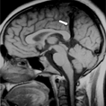 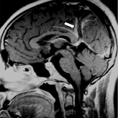
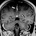 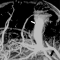 Fig. 1. Persistent falcine sinus without associated anomalies. Precontrast sagittal (A), postcontrast sagittal (B) and postcontrast coronal (C) T1 weighted images showing a falcine sinus in its entire length (white arrow). MR venography (D) showing the falcine sinus (white arrow) and the straight sinus (black arrow). Fig. 1. Persistent falcine sinus without associated anomalies. Precontrast sagittal (A), postcontrast sagittal (B) and postcontrast coronal (C) T1 weighted images showing a falcine sinus in its entire length (white arrow). MR venography (D) showing the falcine sinus (white arrow) and the straight sinus (black arrow).
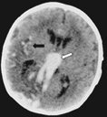  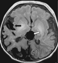
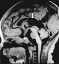 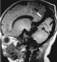 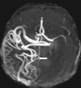 Fig. 2. Persistent falcine sinus associated with aneurysmal dilatation of the vein of Galen and arteriovenous malformation. A: Brain CT showing a markedly dilated vein of Galen (white arrow) and dilated vessels in the right Sylvian fissure (black arrow). B, C, D: Brain MRI showing dilatations of the vein of Galen (white arrow) and arteriovenous malformation (black arrow) in the right hemisphere. The straight sinus is patent and has a terminal stenosis. E: Brain MRI showing a persistent falcine sinus (white arrow) reaching the superior sagittal sinus in its posterior third. The patent straight sinus is associated with terminal stenosis. F: Brain MRA showing arteriovenous malformation (white arrow). Fig. 2. Persistent falcine sinus associated with aneurysmal dilatation of the vein of Galen and arteriovenous malformation. A: Brain CT showing a markedly dilated vein of Galen (white arrow) and dilated vessels in the right Sylvian fissure (black arrow). B, C, D: Brain MRI showing dilatations of the vein of Galen (white arrow) and arteriovenous malformation (black arrow) in the right hemisphere. The straight sinus is patent and has a terminal stenosis. E: Brain MRI showing a persistent falcine sinus (white arrow) reaching the superior sagittal sinus in its posterior third. The patent straight sinus is associated with terminal stenosis. F: Brain MRA showing arteriovenous malformation (white arrow).
Summary
The falcine sinus is a normal intrauterine venous structure located between the dural leaves of the falx cerebri, normally involutes before birth.[1-3] It develops from the mesenchyme in the mesencephalic flexure, the same area that gives rise to the straight sinus.[2] There are a few known conditions associated with a persistent falcine sinus, including malformation of the vein of Galen, arteriovenous malformation, corpus callosum agenesis, bifid cranium, osteogenesis imperfecta, acrocephalosyndactyly (Apert syndrome), Chiari II malformations, absent tentorium, and occipital encephalocele. There are few reported acquired lesions in association with persistent falcine sinus including venous sinus thrombosis or obstruction of the straight sinus by a mass lesion.[4-9] Our first case represents a persistent falcine sinus without associated anomalies, which is an extremely rare condition to our knowledge. The association of aneurysmal dilatation of the vein of Galen and arteriovenous malformation with persistent falcine sinus in our second case has not been reported.
Attention should be paied to the brain to exclude anomalies and also obstruction of the deep venous system in the presence of persistent falcine sinus.
Acknowledgements
The authors want to thank Dr. Chunxiang Wang from Tianjin Children's Hospital, for his assistance in reproduction of photographic images.
Funding: None.
Ethical approval: Not needed.
Competing interest: None declared.
Contributors: Cai CQ wrote the main body of the article under the supervision of Zhang QJ. Shen CH is the guarantor. Yang WD provided advices on medical aspects.
References
1 Strub WM, Leach JL, Tomsick TA. Persistent falcine sinus in an adult: demonstration by MR venography. AJNR Am J Neuroradiol 2005;26:750-751.
2 Sener RN. Association of persistent falcine sinus with different clinicoradiologic conditions: MR imaging and MR angiography. Comput Med Imaging Graph 2000;24:343-348.
3 Manoj KS, Krishnamoorthy T, Thomas B, Kapilamoorthy TR. An incidental persistent falcine sinus with dominant straight sinus and hypoplastic distal superior sagittal sinus. Pediatr Radiol 2006;36:65-67.
4 Kesava PP. Recanalization of the falcine sinus after venous sinus thrombosis. AJNR Am J Neuroradiol 1996;17:1646-1648.
5 Raybaud CA, Strother CM, Hald JK. Aneurysms of the vein of Galen: embryonic considerations and anatomical features relating to the pathogenesis of the malformation. Neuroradiology 1989;31:109-128.
6 Huang YP, Ohta T, Okudera T, Robbins A. Anatomic variations of the dural venous sinuses. In: Kapp JP, Schmidek HH, eds. The cerebral venous system and its disorders. Orlando, Fla: Grune & Stratten, 1984: 109-167.
7 Ratliff J, Voorhies RM. Arteriovenous fistula with associated aneurysms coexisting with dural arteriovenous malformation of the anterior inferior falx. Case report and review of the literature. J Neurosurg 1999;91:303-307.
8 Bartels RH, Merx JL, van Overbeeke JJ. Falcine sinus and occipital encephalocele: a magnetic resonance venography study. J Neurosurg 1998;89:738-741.
9 Kashimura H, Arai H, Ogasawara K, Ogawa A. Persistent falcine sinus associated with obstruction of the superior sagittal sinus caused by meningioma�Dcase report. Neurol Med Chir (Tokyo) 2007;47:83-84.
Received March 5, 2008 Accepted after revision August 18, 2008
|
