|
Esophageal candidiasis in an
immunocompetent girl
Mohammed Yahya Hasosah, Mahmmod Showail, Ashraf Al-sahafi, Mohammed Satti, Kevan Jacobson
Jeddah, Saudi Arabia
Author Affiliations: Department of Pediatrics (Hasosah MY, Showail M, Al-sahafi A) and Department of Pathology (Satti M), King Abdul-Aziz Medical City, National Guard Hospital, Jeddah, Saudi Arabia; Department of Pediatrics, BC Children Hospital, Vancouver, Canada (Jacobson K)
Corresponding Author: Mohammed Yahya Hasosah, Department of Pediatrics, King Abdul-Aziz Medical City, National Guard. PO BOX 8202, Jeddah 21482, Saudi Arabia (Tel: 96626240000; Email: myhasosah@yahoo.com)
doi:10.1007/s12519-009-0031-4
Background: Infections of the esophagus are rare and most commonly seen in children with immune suppression resulting from malignancy, chronic metabolic or infectious disease, or immunosuppressive drug therapy.
Methods: An 18-month-old girl on inhaled corticosteroid for bronchial asthma presented with coffee-ground emesis and melena. Upper endoscopy revealed yellow-white plaques scattered over the mucosa of the distal esophagus. Biopsy results showed chronic esophagitis with features of reflux disease. Gram staining of esophageal brushing showed pseudohyphae, and the culture was positive for candida species.
Results: The patient was treated with omeprazole 2 mg/kg per day and fluconazole 6 mg/kg per day for three weeks. Immunological workup was normal and she was negative for human immunodeficiency virus. Post treatment endoscopy showed normal appearance of esophageal mucosa and normal histology. At 12 months after treatment, the child remained asymptomatic and continued to thrive.
Conclusions: We believe that this child developed esophageal candidiasis secondary to long-term use of inhaled corticosteroid associated with immune suppression on a background of reflux esophagitis. Anti-acid and antifungal therapies are effective in the treatment.
Key words: esophageal candidiasis; reflux esophagitis; upper gastrointestinal bleeding
World J Pediatr 2009;5(2):152-154
Introduction
Infections of the esophagus are rare and most commonly seen in immunocompromised individuals. Common infectious agents include candida, herpes simplex virus and cytomegalovirus.[1] Candida esophagitis occurs mainly in children with immune suppression resulting from malignancy, chronic metabolic or infectious disease, or immunosuppressive drug therapy.[2] The accepted diagnostic criteria for candidal esophagitis include the characteristic endoscopic appearance, microscopic evidence of pseudohyphae in endoscopic mucosal brushings, or invasive candidiasis on biopsy.[3] Herein we present an unusual case of upper gastrointestinal bleeding and food refusal that was ultimately diagnosed as esophageal candidiasis.
Case report
An 18-month-old girl was admitted to our hospital with a history of one-day effortless vomiting associated with coffee-ground emesis and melena. She was refusing solid and liquid food, and appeared to be irritable at swallowing. She was a product of a twin pregnancy, delivered at 34 weeks of gestation with a birth weight of 2.6 kg. She was discharged with her mother after 7 days of observation in the hospital due to prematurity and a ventricular septal defect which closed spontaneously. She was breastfed and supplemented with cow's milk formula feeding to maintain good growth. At the age of 2 months, she was admitted to the hospital for moderately severe gastroenteritis. At the age of 10 months, she was subjected to treatment for respiratory syncytial virus bronchiolitis. Subsequently, she experienced several attacks of wheezing and coughing and was diagnosed with bronchial asthma. Her symptoms responded to a B2-agonist inhaler and fluticasone (FlixotideR) inhaler 50 mg two puffs twice per day. Her growth remained inadequate despite high caloric intake. There was no history of antibiotic use. Her immunizations were up to date and she demonstrated normal development. Three days prior to the current admission, she developed a runny nose, flue-like symptoms associated with fever and cough, which were not responsive to the inhaler therapy. She had no history of abdominal pain, abdominal distension, hematemesis, petechiae or bleeding from any site.
Physical examination revealed that she was conscious, alert, not pale or jaundiced. Her vital signs were stable. Her height was 77 cm (10th centile), weight was 8.2 kg (<10th centile), and head circumference was 43 cm (<10th centile). Her throat was congested and no source of oral or nasal bleeding was evident. Laboratory studies showed hemoglobulin 10.6 g/L, WBC 13.5×10⁹/L, platelets 304 000, and normal serum electrolytes, renal profiles, albumin, total protein, transaminases and coagulation profile. Chest X-ray showed nothing abnormal.
The patient as a case of upper gastrointestinal bleeding was investigated. Upper endoscopy revealed oral thrush and normal duodenal and gastric mucosa. The middle to distal esophagus was covered with white adherent plaques scattered over the mucosa (Fig. 1). The mucosa was hyperemic and friable, and the plaques were adherent. Multiple ulcers were seen in the distal esophagus and there was no active bleeding. The plaques bled easily when brushed for Gram staining and fungal culture. Gram staining showed pseudohyphae and the culture grew candida species. The biopsy results indicated chronic esophagitis with infiltration of inflammatory cells mainly neutrophils and lymphocytes associated with papillary elongation and basal cell hyperplasia. Candida-like organisms with pseudohyphae were also seen (Fig. 2), but there was no tissue invasion.
The child was treated with omeprazole 2 mg/kg per day and intravenous fluconazole 6 mg/kg per day in addition to peripheral total parentral nutrition. Following confirmation of sensitivity to fluconazole, oral fluconazole 6 mg/kg per day was commenced and continued for three weeks. Her emesis and dysphagia to solid food improved gradually.
A thorough investigation was conducted to look for an underlying immunodeficiency. Negative human immunodeficiency virus (HIV) serology and PCR results were found. Levels of serum immunoglobulins were normal for her age. The percentages of CD4 and CD19 cells were normal. T cell subset was within normal limits. Serologically cytomegalovirus and herpes simplex virus were negative.
Two months after the first endoscopy, she underwent another upper endoscopy to confirm mucosal healing and eradication of candida. Upper endoscopy showed normal esophageal mucosa and normal histology. At her most recent follow-up, 12 months after the therapy, she remained asymptomatic and was thriving.
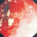
Fig. 1. Endoscopic view showing an edematous and friable esophageal mucosa with white adherent plaques.
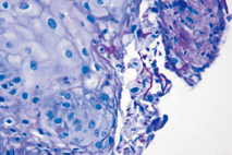
Fig. 2. Budding filaments and pseudohyphae consistent with Candida species (arrows), admixed with detached fragments of squamous epithelium and lymphocytes (PAS stain, original magnification × 400).
Discussion
In patients at risk for candidiasis, candida colonization of mucosal surfaces is the first pathologic stage, followed by epithelial and sometimes deeper invasion by pseudohyphae.[4] Risk factors for esophageal candidiasis include malignancy, chronic metabolic or infectious disease, or immunosuppressive drug therapy. Other predisposing factors include broad spectrum antibiotic therapy, corticosteroids, diabetes, blood dyscrasias and HIV infection.[5]
In candida esophagitis, the esophageal mucosa is typically friable and erythematous with ulcers covered by thick adherent white exudates in contrast to herpetic esophagitis.[6] Because the plaques in the esophagus could not be washed off, they were unlikely swallowed oral thrush lesions. Moreover the esophageal plaques bled easily when brushed, supporting their intrinsic esophageal origin.
In patients with candida esophagitis, the most common complaints are difficulty and pain with swallowing. Our patient had upper gastrointestinal bleeding and food refusal. The cause of these symptoms is likely due to the combination of esophageal injury associated with candida infection and acid reflux. While the upper gastrointestinal bleeding is an unusual manifestation of esophageal candidiasis, acid reflux associated esophageal mucosal ulceration is commonly manifested with upper gastrointestinal bleeding.
Long-term use of high-dose corticosteroids suppresses lymphocyte and granulocyte function and predisposes the patient to mucosal infection with candida.[7] Underwood et al[8] retrospectively reviewed 18 patients with endoscopically proven candida esophagitis and found that 4 of these 18 patients were using inhaled steroid. We believe that the inhaled steroid suppressed the immune function of our patient. It is likely that her candida infection was related to an improper long-term use of inhaled steroid over 8 months before admission.
Antifungal agents used in esophageal candidiasis treatment include nystatin, fluconazole, flucytosine, itraconazole and amphotericin B.[9] Nystatin is less efficacious in immunosuppressed patients. Fluconazole therapy for 14 days is usually well tolerated and effective for esophageal candidiasis.
In summary, we described a girl with no congenital or acquired immunodeficiency who had esophageal candidiasis superimposed on reflux esophagitis.[10-12] We found that candida esophagitis was secondary to the complication of inhaled steroid or esophageal dysmotility of reflux esophagitis or both. The patient responded well to anti-acid and antifungal therapies.
Funding: None.
Ethical approval: Not needed.
Competing interest: No benefits in any form have been received or will be received from any commercial party related directly or indirectly to the subject of this article.
Contributors: Hasosah MY wrote the article and is the guarantor. All authors approved the final version of the paper.
References
1 McDonald GB, Sharma P, Hackman RC, Meyers JD, Thomas ED. Esophageal infections in immunosuppressed patients after marrow transplantation. Gastroenterology 1985;88:1111-1117.
2 Misra S, Ament ME. Esophagitis. In: Feign RD, Cherry JD, eds. Textbook of Pediatric Infectious Diseases. Philadelphia: WB Saunders, 1997: 562-566.
3 López-Dupla M, Mora Sanz P, Pintado García V, Valencia Ortega E, Uriol PL, Khamashta MA, et al. Clinical, endoscopic, immunologic, and therapeutic aspects of oropharyngeal and esophageal candidiasis in HIV-infected patients: a survey of 114 cases. Am J Gastroenterol 1992;87:1771-1776.
4 Walsh TJ, Pizzo PA. Nosocomial fungal infections: a classification for hospital-acquired fungal infections and mycoses arising from endogenous flora or reactivation. Annu Rev Microbiol 1988;42:517-545.
5 Chocarro Martínez A, Galindo Tobal F, Ruiz-Irastorza G, González López A, Alvarez Navia F, Ochoa Sangrador C, et al. Risk factors for esophageal candidiasis. Eur J Clin Microbiol Infect Dis 2000;19:96-100.
6 Yacono JV. Type I herpes simplex esophagitis with candidal esophagitis in an immunocompetent host. N Y State J Med 1985;85:656-658.
7 Mathieson R, Dutta SK. Candida esophagitis. Dig Dis Sci 1983;28:365-367.
8 Underwood JA, Williams JW, Keate RF. Clinical findings and risk factors for Candida esophagitis in outpatients. Dis Esophagus 2003;16:66-69.
9 Ostrosky-Zeichner L, Rex J, Bennett J. Deeply invasive candidiasis. Infect Dis Clin North Am 2002;16:821-835.
10 Billings CJ, Billings AM, Crissinger KD, Gremse DA. Candida esophagitis in infants with gastresophageal reflux disease and feeding intolerance. Am J Gastroenterol 2004;99 Suppl:S315.
11 Uc A, North P, Burks W. Esophageal candidiasis in an infant with reflux esophagitis. J Pediatr Gastroenterol Nutr 2000;31:572-574.
12 Rahhal RM, Ramkumar DP, Pashankar DS. Simultaneous herpetic and candidal esophagitis in an immunocompetent teenager. J Pediatr Gastroenterol Nutr 2005;40:371-373.
Received May 27, 2008 Accepted after revision August 26, 2008
|
