| |
Duodenal heterotopic pancreas in a child
Li-Duan Zheng, Qiang-Song Tong, Shao-Tao Tang, Zhi-Yong Du, Qing-Lan Ruan
Wuhan, China
Author Affiliations: Department of Pathology, Union Hospital of Tongji Medical College, Huazhong University of Science and Technology, Wuhan 430022, China (Zheng LD); Department of Pediatric Surgery, Union Hospital of Tongji Medical College, Huazhong University of Science and Technology, Wuhan 430022, China (Tong QS, Tang ST, Du ZY, Ruan QL). Zheng LD and Tong QS contributed equally to this work
Corresponding Author: Qiang-Song Tong, Department of Pediatric Surgery, Union Hospital of Tongji Medical College, Huazhong University of Science and Technology, Wuhan 430022, China (Tel: 86-27-63776478; Email: qs_tong@hotmail.com)
doi:10.1007/s12519-009-0029-y
Background: Heterotopic pancreas is characterized by pancreatic tissue outside the pancreatic bed. However, duodenal heterotopic pancreas in children is rarely reported so far. We describe herein duodenal heterotopic pancreas in a child who suffered from chronic abdominal pain.
Methods: An 8-year-old boy presented with upper abdominal pain and intermittent vomiting, without a history of melena, hematochezia, hematemesis, clay-colored stools, jaundice, hepatitis or dyscrasia. No specific medication or change in position relieved the pain. Based on the elevated serum amylase levels, and the findings of CT, barium meal X-ray examination, magnetic resonance imaging, and upper gastrointestinal endoscopy, a duodenal mass was diagnosed initiatively. Intraoperative frozen section analysis was performed for the diagnosis. The mass was dissected.
Results: Intraoperative frozen section analysis and routine pathological examination confirmed the diagnosis of duodenal heterotopic pancreas. The patient had an uneventful recovery and remained asymptomatic postoperatively during a follow-up period of 16 months.
Conclusions: Heterotopic pancreas should be considered in children with a duodenal mass and abdominal pain. Intraoperative frozen section analysis is helpful in the diagnosis of the disease. Surgical treatment of the lesion should be performed to prevent bleeding, ulceration, outlet obstruction or malignant degeneration.
Key words: child; duodenum; heterotopic pancreas
World J Pediatr 2009;5(2):146-148
Introduction
Heterotopic pancreas is found in 1% to 2% of autopsies and 0.5% of upper abdominal laparotomies.[1] This anomaly may involve any segment of the digestive tract, most frequently in the stomach, duodenum, or jejunum, and more rarely in Meckel's diverticulum, the papilla of Vater, the gallbladder, and the umbilicus.[1] Although ectopic pancreas is most often noted as an incidental finding at autopsy, it is capable of producing symptoms such as bleeding, ulceration, outlet obstruction, pancreatitis and even malignant degeneration, depending on its location, size, and involvement of the overlying mucosa.[2] Pancreatic heterotopy can be the site of tumor development from one of the cellular elements of heterotopy, or predispose to the formation of small cysts surrounded by inflammation and scarring.[3] This phlogistic pattern of presentation is called the cystic dystrophy of heterotopic pancreas, which can be associated with chronic pancreatitis of the true pancreas.[3] However, duodenal heterotopic pancreas has been rarely reported in children so far. This report describes one case of duodenal heterotopic pancreas that caused chronic abdominal pain in a child.
Case report
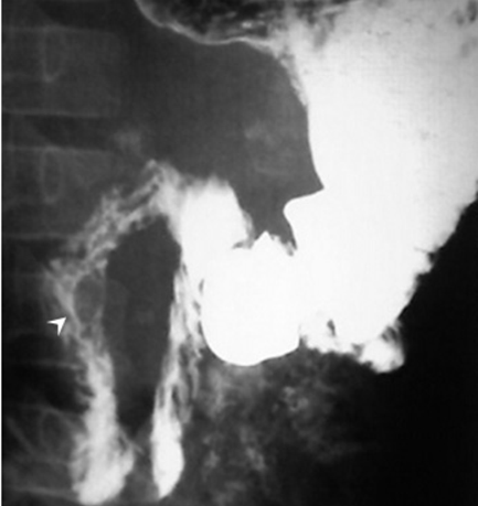
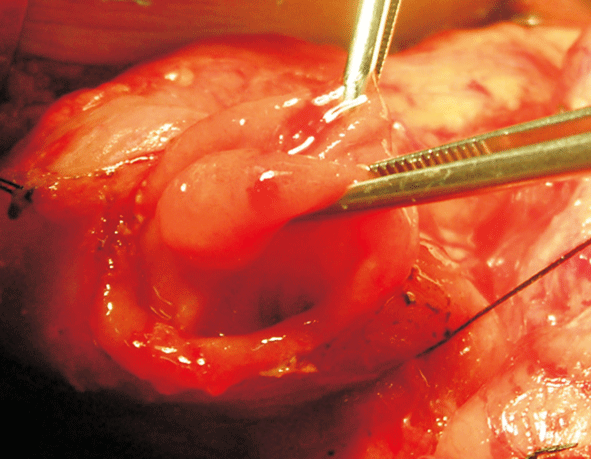
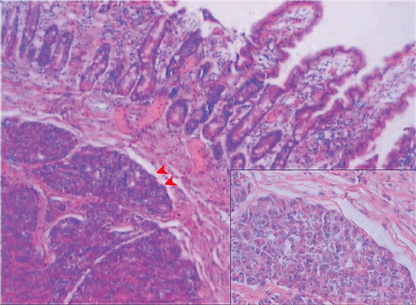
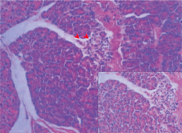
An 8-year-old boy was admitted to our hospital because of severe upper abdominal pain and intermittent vomiting in the preceding 12-month period. There was no history of melena, hematochezia, hematemesis, clay-colored stools, jaundice, or hepatitis, and he did not describe any food dyscrasias although fatty food seemed to make the symptoms worse. No specific medication or change in position relieved the pain. One week before admission, the patient undertook CT in another hospital, which indicated normal liver, spleen and pancreas. On admission, physical examination revealed mild tenderness in the upper abdomen. Hematological and blood biochemical tests showed a 4-fold increase in the level of serum amylase (365 U/L, reference range 20-90 U/L) accompanied with an elevated level of alkaline phosphatase, but markers of acute hepatocyte injury such as alanine, aspartate aminotransferase, and gamma glytamyl transpeptidase (GT) and bilirubin levels remained normal. Acute pancreatitis was initially suspected. Ultrasonographic examination revealed normal liver, no stones in the gallbladder, and a common bile duct with a diameter of 5 mm and with no signs of stone obstruction. The second portion of duodenal wall appeared thicker and a small mass (0.9 cm in diameter) was present. The head of the pancreas appeared normal. A barium meal revealed a small filling defect of the second portion of the duodenum (Fig. 1). Magnetic resonance imaging results showed a 0.9 cm ¡Á 1.0 cm solid tumor in the descending duodenum. Upper gastrointestinal endoscopy also revealed a severe semispherical deformation at the second portion of the duodenum, without evidence of mucosal change or central umbilication. Based on these examinations, a duodenal mass was suspected. Since the parents refused the endoscopic ultrasonography with linear biopsy, intraoperative frozen section analysis was performed because upper abdominal pain was severe and malignant tumor was suspected. Histological analysis indicated that the mass was not connected with the pancreatic head, and the proximal margin of the mass was 2 cm distal to the papilla of Vater. Intraoperative frozen section analysis indicated no tumor cells in the heterotopic pancreas. Therefore, the mass was resected. Macroscopically, the specimen of the recected mass was light yellow in color, palpated as firm as tendon tissue, located in the submucosa propria of thickened duodenal wall measuring 1.0¡Á1.0¡Á0.9 cm3 (Fig. 2). Pathological examinations revealed the acinar cells (Fig. 3A), duct cells (Fig. 3A), and islets of Langerhans (Fig. 3B), which confirmed a diagnosis of submucosal heterotopic pancreas of the duodenum. The patient had an uneventful recovery and remains asymptomatic postoperatively during a follow-up period of 16 months.

Fig. 1. Barium meal for the upper gastrointestinal tract. Radiologic examination demonstrated a small filling defect of the second portion of the duodenum and a marked mass located at the descending part of the duodenum.

Fig. 2. Surgical specimen of the resected heterotopic pancreas of the duodenum. There was no erosion or ulcer in the duodenal mucosa. The resected duodenal specimen showed a yellow and round mass, measuring 1.0¡Á1.0¡Á0.9 cm3.
 
Fig. 3. Histological examination of surgical specimen. A: submucosal heterotopic pancreas of the duodenum with acinar cells, duct cells, and islets of Langerhans (HE, original magnification ¡Á 100) (Arrows show islet of acinar cells); B: marked islets of Langerhans between the mucosal and muscular layers of the duodenum (HE, original magnification ¡Á 200) (Arrow shows islet of Langerhans)
Discussion
Heterotopic pancreas is defined as pancreatic tissue outside the pancreatic bed with no anatomic or vascular connection to the pancreas.[1] Despite its congenital origin, pancreatic heterotopia was usually discovered in adults due to its complications.[4] Heterotopic pancreas is usually asymptomatic; however, sometimes there are evident clinical symptoms. The most common symptoms attributed to heterotopic pancreas are abdominal pain, nausea, vomiting, anemia, weight loss and melena.[5] In this case, we observed some lymphocytes surrounding the heterotopic pancreas in the submucosal layer of the duodenum, suggesting chronic inflammation. In addition, the serum amylase of this patient was 4-fold more than normal level. We suspected that abdominal pain might be due to ectopic pancreatitis.
Despite many advances of new diagnostic tools and methods, the diagnosis of heterotopic pancreas prior to surgery is still difficult.[5] It is known that endoscopy is helpful in diagnosis of heterotopic pancreas located in the submucosa or muscular layer of the stomach and in the first and second portions of the duodenum.[5] Endoscopic ultrasonography is a reliable method providing precise localization, extent and characteristics of the submucosal mass, making it possible to differentiate tumor from pancreas annulare.[6] In addition, surgical findings and intraoperative histology play important roles in making an accurate diagnosis. In this case, because of the disagreement of parents on endoscopic ultrasonography with linear biopsy, intraoperative biopsy was critical for the diagnosis. According to Heinrich, heterotopic pancreatic tissue can be divided histologically into three types: type I, normal pancreas in the arrangement of their lobules, acini, and ducts; type II, imperfect lobules or no lobule arrangement; and type III, only ducts are present.[7] In this case, type I heterotopic pancreas of the duodenum was shown by routine hematoxylin & eosin (HE) staining.
Surgical treatment is usually recommended for symptomatic patients with heterotopic pancreas. Conservative surgical procedures including segmental duodenal resection could be an alternative approach to the Whipple procedure.[8] Radical resection which may increase the potential risk of intraoperative complication is not advisable unless cancer has developed.[9] In this case, heterotopic pancreas was not associated with bleeding, ulceration, outlet obstruction or malignant degeneration, surgical resection of the mass was feasible and effective because the 16-month follow-up indicated that the patient had an uneventful recovery and remained asymptomatic postoperatively.
Funding: This work was supported by the grants from the National Natural Science Foundation of China (No. 30200284, No. 30600278 and No. 30772359) and the Program for New Century Excellent Talents in University (NCET-06-0641).
Ethical approval: Not needed.
Competing interest: No benefits in any form have been received or will be received from a commercial party related directly or indirectly to the subject of this article.
Contributors: Tong QS wrote the article. Zheng LD performed the pathological examinations. All authors were responsible for the follow-up of treatment protocol. Tong QS is the guarantor.
References
1 Chou SJ, Chou YW, Jan HC, Chen VT, Chen TH. Ectopic pancreas in the ampulla of vater with obstructive jaundice. A case report and review of literature. Dig Surg 2006;23:262-264.
2 Ueno S, Ishida H, Hayashi A, Kamagata S, Morikawa M. Heterotopic pancreas as a rare cause of gastrointestinal hemorrhage in the newborn: report of a case. Surg Today 1993;23:269-272.
3 Indinnimeo M, Cicchini C, Stazi A, Ghini C, Laghi A, Memeo L, et al. Duodenal pancreatic heterotopy diagnosed by magnetic resonance cholangiopancreatography: report of a case. Surg Today 2001;31:928-931.
4 Lai EC, Tompkins RK. Heterotopic pancreas. Review of a 26 year experience. Am J Surg 1986;151:697-700.
5 Hsia CY, Wu CW, Lui WY. Heterotopic pancreas: a difficult diagnosis. J Clin Gastroenterol 1999;28:144-147.
6 Tison C, Regenet N, Meurette G, Miralli¨¦ E, Cassagnau E, Frampas E, et al. Cystic dystrophy of the duodenal wall developing in heterotopic pancreas: report of 9 cases. Pancreas 2007;34:152-156.
7 Heinrich H. The histology of a contributory tissue. Accessory pancreas. Virchows Arch Pathol Anat 1909;198:392-401.
8 Shi H, Zhang Q, Teng H, Chen JC. Heterotopic pancreas: report of 7 patients. HBPD Int 2002;1:299-301.
9 Connolly MM, Dawson PJ, Michelassi F, Moossa AR, Lowenstein F. Survival in 1001 patients with carcinoma of the pancreas. Ann Surg 1987;206:366-372.
Received April 15, 2008 Accepted after revision June 29, 2008
|

