|
Eosinophilic cystitis in nine children
Ming Liu, Yu-Zhen Zhang, Yu-Hua Li, Hua Xie
Shanghai, China
Author Affiliations: Department of Radiology, Xin Hua Hospital Affiliated to Shanghai Jiaotong University, Shanghai 200092, China (Liu M, Zhang YZ, Li YH); Department of Radiology, Xin Hua Hospital Affiliated to Shanghai Jiaotong University, Shanghai 200092, China (Xie H)
Corresponding Author: Ming Liu, MD, Xin Hua Hospital Affiliated to Shanghai Jiaotong University, Shanghai 200092, China (Tel: 86-21- 65790000 ext 5003; Email: lesserniuniu@126.com)
Background: We described the clinical and imaging manifestations of eosinophilic cystitis and eosinophilic granulomatous cystitis in children.
Methods: The clinical and imaging (mainly CT) manifestations of 7 patients with pathologically proved eosinophilic cystitis and 2 patients with eosinophilic granulomatous cystitis were analyzed retrospectively after cystoscopic tissue biopsy or surgery.
Results: Six boys and three girls, aged 3-13 years (mean 8.3 years), complained of hematuria, irritative voiding, dysuria and abdominal pain. CT scans showed localized or nodular lesions in 4 patients, 2 of them had proved eosinophilic granulomatous cystitis, and others had diffused lesions. All patients were subjected to bladder autoaugmentation, mass resection or medication with corticosteroids and antibiotics.
Conclusions: The clinical and imaging (mainly CT) manifestations of eosinophilic cystitis and eosinophilic granulomatous cystitis in children are apt to be confused with other bladder tumors. Biopsy is necessary for confirming the diagnosis of the lesion and to select the proper management.
Key words: bladder; tumor; cystitis; eosinophils; biopsy; computed tomography
World J Pediatr 2007;3(3):222-225
Introduction
Benign bladder tumors with eosinophilic infiltration are rare in children and often misdiagnosed as malignant lesions even by experienced pathologists. From 2001 to 2004, we diagnosed and treated 7 patients with eosinophilic cystitis and 2 patients with eosinophilic granulomatous cystitis. We reviewed their clinical manifestations and computed tomography (CT) findings in addition to their diagnosis and management.
Clinical summary
Medical records, imaging, and pathological examina-tions (tissue biopsy or surgery) of the 9 patients showed pathologically proved eosinophilic cystitis (7 patients) and eosinophilic granulomatous cystitis (2 patients). The literature on inflammatory eosinophilic bladder tumors as well as their clinicopathological and imaging features and treatment in children was reviewed.
In our patients, unenhanced and contrast-enhanced CT scan was used to evaluate the lesions, by a GE lightspeed scanner (thickness 5.0-7.5 mm, interval 7.5-15 mm, Omnipaque 300 mgI/ml, and dosage 1.0-1.5 ml/kg).
Symptoms, clinical findings, imaging features and treatment of the 9 patients are shown in the Table.
Table. Presentations, diagnosis and treatment of eosinophilic cystitis and eosinophilic granulomatous cystitis in the nine patients
|
Age (y) |
Sex |
Hematuria |
Irritative voiding |
Dysuria |
Abdominal pain |
CT findings of the bladder wall |
Treatment |
|
7* |
F |
+ |
- |
- |
+ |
Nodular and sessile mass |
Mass resection |
|
5 |
F |
- |
+ |
- |
- |
Diffused thickened wall |
Autoaugmentation |
|
10 |
M |
- |
- |
+ |
+ |
Diffused thickened wall |
Lost to follow-up |
|
3 |
M |
- |
+ |
+ |
- |
Diffused thickened wall |
Conservative |
|
13 |
M |
+ |
- |
+ |
- |
Nodular and sessile mass |
Mass resection |
|
11 |
M |
- |
+ |
- |
- |
Diffused thickened wall |
Conservative |
|
8† |
M |
- |
+ |
- |
- |
Locally thickened right wall |
Conservative |
|
9 |
M |
+ |
- |
+ |
- |
Diffused thickened wall |
Conservative |
|
9* |
F |
- |
+ |
- |
+ |
Thickened left-posterolateral wall with
a mass protruding to the pelvis |
Mass resection |
One patient with eosinophilic granulomatous cystitis had intermittent abdominal pain for 3 years and gross hematuria for 4 days, other patients complained of the pain lasting from 5 days to 6 months. None of the 9 patients had urinary tract infection at presentation or evident pathogenic allergens.
Ultrasonography and computerized tomography of the pelvis showed a focal mass of the bladder or locally or diffusely thickened bladder walls in the 9 patients (Figs. 1-5).
A B B C
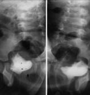
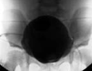
Fig. 1. Eosinophilic cystitis. A: Contrast-enhanced CT showed diffused thickened and marked enhanced bladder wall. B: Cystogram showed only 40 ml of bladder capacity, with irregular-defined margin. C: Postoperative cystogram revealed 250 ml of bladder capacity, well-defined margin following autoaugmentation after 9-month follow-up.
A B C
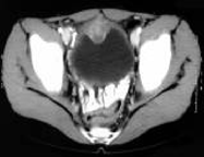
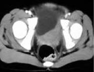
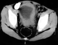
Fig. 2. Eosinophilic cystitis. A: Unenhanced CT showed a nodular and sessile mass along the bladder dome. B: Contrast-enhanced CT showed the mass with heterogeneously moderate-enhanced. C: Ultrasound also showed the mass clearly.
A B C
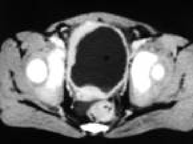
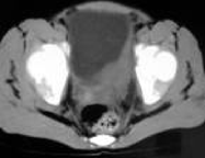
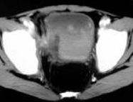
Fig. 3. Eosinophilic granulomatous cystitis. A&B: CT showed the left posterolateral thickened bladder wall accompanied with a large marked firm mass protruding to pelvis, and with moderate-enhanced. C: Nothing was found in cystogram.
A B C
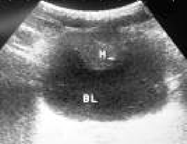
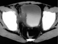
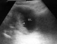
Fig. 4. Eosinophilic cystitis. A: CT and ultrasound showed the right lateral thickened bladder wall. B: At contrast-enhanced CT the lesion appeared slightly-enhanced. C: Ultrasound showed the lesions more clearly.
A B
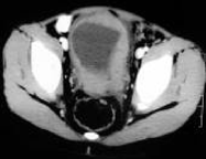
Fig. 5. Eosinophilic cystitis. A&B: CT showed diffusely irregularly thickened bladder wall with homogeneously and slightly-enhanced.
Urography revealed a small contracted bladder with diffused lesions, but without appearance of hydronephrosis. Cystoscopy and diagnostic biopsy revealed erythematous changes in all patients. Additional findings included a tumor-like mass in 2 patients, and diffusely edematous change in 3. In each patient, the histological changes of eosinophilic cystitis, namely some combination of diffuse eosionphilic inflammation throughout the lamina propria and muscularis accompanied by muscle necrosis and submucosal edema, were identified by biopsy. In the two patients with proved eosinophilic granulomatous cystitis, despite the presence of eosinophils, multinucleate giant cells were observed histologically.
One patient was lost to follow-up after cystoscopic biopsy, and 7 patients showed improvement after mass resection or oral administration of corticosteroids and antibiotics. They were followed up for 5 weeks to 28 months. The last patient, a 5-year-old girl, showed symptoms such as irritation during the prolonged administration of steroids and antibiotics. Finally she underwent bladder autoaugmentation. Urodynamic studies before and after operation showed marked improvement and the capacity of the poorly compliant bladder increased from 40 ml to 250 ml within 9 months of follow-up. None of the 8 patients had recurrence during follow-up.
Discussion
Inflammatory eosinophilic bladder tumor is rare in children. But as one of the benign bladder lesions, the tumor has received more attention from pediatricians. Since its initial description,[1,2] only less than 40 anecdotal cases in children have been reported.[3-12] Eosinophilic cystitis refers to diffuse eosinophilic bladder infiltration or an inflammatory eosinophilic tumor.[11] The cause of the disease remains unclear, but allergic reaction is recognized as the etiology.[13,14] IgE may bind to various antigens, activating mast cell degranulation, eosinophil attraction and release of damaging enzymes. Pathologically, there is predominant eosionphilic infiltration throughout the lamina propria and muscularis. Submucosal edema, muscle necrosis, and superficial muscle fibrosis may occur. Severe lesions are associated with mucosal ulceration. Histological examination revealed multinucleate giant cells, which indicate the appearance of eosinophilic granulomatous cystitis.[6,10] The allergic reaction may be due to bacteria, food, parasites, medication, and others. Diffuse lesions are induced by allergens from food or drugs whereas local lesions from parasites.[14] Because there are no cause-and-effect relationships, the eosinophilic inflammatory response may be a common manifestation of several etiological pathways. In our series, allergic reaction was noted in only one patient who had taken drug for three years.
Inflammatory eosinophilic bladder tumor was confirmed with a male predilection. Eosinophilic cystitis affects boys more often than girls, with a ratio of about 2:1 and a mean age of 6 years in children.[4,15] Although a few asymptomatic cases have been reported, most of them present with hematuria, irritative voiding, dysuria and abdominal pain in combination.[4,13-15] In our series, one patient with eosinophilic granulomatous cystitis had an intermittent abdominal pain for 3 years and gross hematuria for 4 days, and other patients had the hematuria for 5 days to 6 months without any special manifestations. Generally, more chronic lesions suggest the appearance of a granulomatous process and the duration of hematuria in most patients may last weeks to months. In our series, however, the patient with eosinophilic granulomatous cystitis only had irritative voiding for 5 days. It was reported in some cases that peripheral eosinophil count exceeding 5% of the total leukocyte count or reaching 500 per ml blood.[16] In our series, an 8-year-old boy had peripheral eosinophils accounting for 16% of the total leukocyte count. Impaired renal function was attributed to unilateral or bilateral hydronephrosis caused by eosinophilic infiltration and later fibrosis of the ureters in chronic late-stage cases. Urography revealed abnormalities or diffused lesions in the contracted bladder but without hydronephrosis.
Cystoscopic abnormalities comprise a wide spectrum of inflammatory bladder tumors with eosinophilic infiltration, which vary from mucosal erythema to raised velvety, polypoid, edematous and frankly fungating, and invasive-appeared masses,[12,13] Thus, eosinophilic lesions may present as a diffused eosinophilic inflammation of the bladder wall without a mass or as a nodular or sessile lesion on computerized tomography and ultrasonography.[17] Eosinophilic cystitis should be referred to infiltrative and global cystitis without a mass, and the mass should be considered as inflammatory eosinophilic tumor.[6] In our opinion, they are inflammatory eosinophilic bladder tumors including eosinophilic granulomatous cystitis (usually with a mass) and eosinophilic cystitis (with or without a mass) because the thickened bladder wall secondary to eosinophilic infiltration appears to be asymmetrical and irregular in some tumor-like regions. Ultrasonography and CT of the pelvis showed a papillary and sessile mass along the bladder dome in 2 patients (22%) of our series with eosinophilic cystitis and eosinophilic granulomatous cystitis respectively. The imaging characteristics of the two patients could mimic each other, but histologically multinucleate giant cells were observed in eosinophilic granulomatous cystitis. There were some CT characteristics in inflammatory eosinophilic bladder tumors. Unenhanced CT showed that the lesion appeared as a homogeneous density with isoattenuation relative to soft tissues. Contrast-enhanced CT showed that the local lesion appeared to be heterogeneous and the diffused lesion appeared to be homogeneous. The enhancement varied, but moderate enhancement predominated. The diffused thickened bladder wall with moderate enhancement was characteristic in eosinophilic cystitis. In our series, the ratio was 5/9 (55.6%) or 5/7 (71.4%).
The association of eosinophilic cystitis with chronic granulomatous disease (CGD) has been reported in less than 10 patients.[5,10,18] As a primary immunodeficiency, CGD affects the oxidative mechanism of microbial killing of phagocytic cells. The patients with CGD also develop gastrointestinal and genitourinary obstructive granuloma. Although eosinophilic cystitis has a benign course in children, CGD can be recurrent and might be dependent on therapy with corticosteroids. Eosinophilic granulomatous cystitis is characterized by multinucleate giant cells, but not by progressive eosinophilic cystitis.
The clinical and imaging manifestations of inflammatory eosinophilic bladder tumors are difficult to distinguish from those of such malignant tumors as sarcoma and others like inflammatory myofibroblastic tumors. Histological examination showed that eosinophils may be present in the inflammatory myofibroblastic tumors but they are not prominent. Since myofibroblastic tumor is considered benign but tends to be locally invasive, open surgery is required with bladder preservation.[6] Inflammatory eosinophilic tumors can be treated with antihistamine agents and steroids, in addition to surgical intervention.[5,9,11-13,15] The diagnosis of inflammatory eosinophilic tumors is based on histological examination. Tissue biopsy is necessary to confirm the diagnosis of bladder lesions and to select proper management.
Funding: None.
Ethical approval: Not needed.
Competing interest: None declared.
Contributors: LM proposed the study and wrote the main body of this paper. ZYZ and LYH contributed to the design and interpretation of the study and provided advice on medical aspect. XH collected the data and clinical manifestations of the patients.
References
1 Brown EW. Eosinophilic granuloma of the bladder. J Urol 1960;83:665-668.
2 Palubinskas AJ. Eosinophilic cystitis. Case report of eosinophilic infiltration of the urinary bladder. Radiology 1960;75:589-591.
3 Redman JF, Parham DM. Extensive inflammatory eosinophilic bladder tumors in children: experience with three cases. South Med J 2002;95:1050-1052.
4 Sujka SK, Fisher JE, Greenfield SP. Eosinophilic cystitis in children. Urology 1992;40:262-264.
5 Ladocsi LT, Sullivan B, Hanna MK. Eosinophilic granulomatous cystitis in children. Urology 1995;46:732-735.
6 Netto JM, Perez LM, Kelly DR, Joseph DB. Pediatric inflammatory bladder tumors: myofibroblastic and eosinophilic subtypes. J Urol 1999;162:1424-1429.
7 Verhagen PC, Nikkels PG, de Jong TP. Eosinophilic cystitis. Arch Dis Child 2001;84:344-346.
8 Pomeranz A, Eliakim A, Uziel Y, Gottesman G, Rathaus V, Zehavi T, etc. Eosinophilic cystitis in a 4-year-old boy: successful long-term treatment with cyclosporin A. Pediatrics 2001;108:E113.
9 Axelrod SL, Ring KS, Collins MH, Reiley EA, Hensle TW. Eosinophilic cystitis in children. Urology 1991;37:549-552.
10 Bauer SB, Kogan SJ. Vesical manifestations of chronic granulomatous disease in children. Its relation to eosinophilic cystitis. Urology 1991;37:463-466.
11 Gerharz EW, Grueber M, Melekos MD, Weingaertner K, Barth P, Riedmiller H. Tumor-forming eosinophilic cystitis in children. Case report and review of literature. Eur Urol 1994; 25:138-141.
12 Sutphin M, Middleton AW Jr. Eosinophilic cystitis in children: a self-limited process. J Urol 1984;132:117-119.
13 Itano NM, Malek RS. Eosinophilic cystitis in adults. J Urol 2001;165:805-807.
14 Littleton RH, Farah RN, Cerny JC. Eosinophilic cystitis: an uncommon form of cystitis. J Urol 1982;127:132-133.
15 van den Ouden D. Diagnosis and management of eosinophilic cystitis: a pooled analysis of 135 cases. Eur Urol 2000;37:386-394.
16 Goldstein M. Eosinophilic cystitis. J Urol 1971;106:854-857.
17 Hansen MV, Kristensen PB. Eosinophilic cystitis simulating invasive bladder carcinoma. Scand J Urol Nephrol 1993;27: 275-277.
18 Barese CN, Podesta M, Litvak E, Villa M, Rivas EM. Recurrent eosinophilic cystitis in a child with chronic granulomatous disease. J Pediatr Hematol Oncol 2004;26:209-212.
Received March 12, 2007 Accepted after revision April 28, 2007
|
